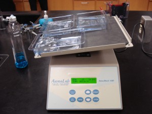
Comparative Proteomics Kits I and II purchased from:

Share in the excitement by watching the newly-released Western Blot music video!
View this Western Blot animation for an overview of the blotting process.
Lesson 1: Protein Extraction From Muscle
Muscle proteins were extracted from five species of fish: flounder, salmon, grouper, rainbow trout, and swordfish.


The four main ingredients in Laemmli buffer are Tris, SDS, bromophenol blue, and glycerol. What is the purpose of each component?
After placing the fish muscle samples in Laemmli buffer, you boiled the samples. What is the purpose of boiling the samples?
Lesson 2: Separate Proteins by SDS-PAGE
What is the purpose of the prestained (Kaleidoscope) standards?
What is the purpose of the actin and myosin standards?
What is the purpose of running the fish muscle proteins in a gel?
Notice all of the fish proteins in lanes 4-8 of the stained polyacrylamide gel:
Lesson 3: Perform Western Blotting
Prior to performing the Western Blot, why did we equilibrate the gels in blotting buffer?
When building the “sandwich” it was imperative to place the gel on blotting paper. Why?
What is the purpose of the nitrocellulose membrane (it’s placed on top of the gel in the “sandwich”)?
In this picture, you’ll notice a student gently rolling the membrane to prevent air bubbles. Why must there be a tight seal between the gel and the membrane?
What is the goal of blotting? Why is an ice-pack placed in the Tetra tank?
Observe the nitrocellulose membrane carefully. Although they are difficult to see, notice the Kaleidoscope (prestained) standards have been transferred to the membrane from an unstained gel. What does this suggest about the transfer of fish muscle proteins to the membrane?
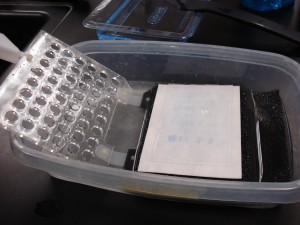
Observe the nitrocellulose membrane that was blotted from a stained gel. Were the fish muscle proteins successfully transferred to the membrane?
Lesson 4: Immunodetection
After completing the blot, why was the membrane placed in blocking solution? (Hint: A major component of the blocking solution is milk).
After the membranes have incubated in blocking solution for at least 15 minutes, you added primary antibody to the membrane. Why?
After allowing the membrane to incubate with the primary antibody for 10 minutes, why did you wash the membrane?
Next, you added secondary antibody to the membrane. Why?
After incubating the membrane with secondary antibodies, you washed it and then added HRP substrate. What is the purpose of the substrate?
Observe the nitrocellulose membrane below. Where is the myosin light chain 1 located in each sample?


